Israeli researchers have identified the biological processes that happen specifically in severe cases of COVID-19 when compared to mild cases. This was done by studying the activity in single infected cells, while the study headed by Prof Ido Amit is from the Department of Immunology at the Weizmann Institute of Science in Rehovot.
Cells in the lungs of severely ill patients called macrophages that usually help in getting rid of lung infection get replaced by cells that increase their illness, found the research. Further finding that coronavirus neutralizes T-cells which fights the infection as an immune response. This allows viruses to damage the body, as published in the journal Cell on Friday.
Chinese researcher also collaborated in the research. This research prompts finding 'weak spots' when a coronavirus patient deteriorates. This paves way for more effective treatments preventing deaths and severity of cases.
Picture of cell used in the study

It is already known that after a week of mild symptoms if there is any severity of COVID-19 due to the highly active immune response called 'cytokine storm' often leads to serious damages such as multiple organ failure especially heart, liver and kidneys.
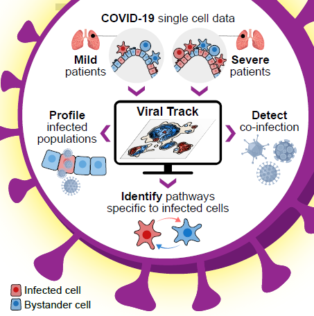
Obtaining a picture of the cell in a given time led to comparing the differences between the activity of cells attacked by SARS-CoV-2 in severely and mildly infected patients. This further led scientists in seeing those cells and genes getting activated, simultaneously knowing which gets silenced, this allowed how inter-cellular communication changes.

The research finds that severe cases of COVID-19 saw damage to macrophage cells, which clears the infection in the lungs. This was done using the state-of-the-art genomic technologies that include something called 'single-cell genomics' developed and led by Prof Amit, according to a report in Haaretz.
Mapping cell activity
The technology can precisely map cellular activity at any moment, by genetic sequencing of the entire viral RNA. This means mapping can be done after the coronavirus invades a cell and starts multiplying.
In doing so, this study analyzed almost a million cells taken from the lung fluid of seriously ill patients, mildly ill and of the healthy. The mapping found that the novel coronavirus usually attacked epithelial cells, responsible for respiration in the lungs. After the infection, the "immune environment" of lungs gets totally transformed, explained Amit.
In severe cases, macrophages in the lungs are replaced by immune system cells such as monocytes and blood cells accelerating the cytokine storm in which the T-cells which play an important role in healing the inflammation become neutralized and inactive.
This change also causes indirect damages like getting infected by other viruses which the body had managed to repulse. This explains why people with existing conditions are more prone to danger. This has prompted in developing clinical studies that determine the right treatments to protect macrophages.


