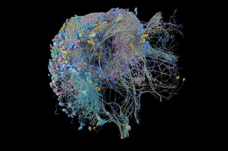
When it comes to imagining a brain, most of the people picture an organ with several wrinkles, while for medical students and doctors it is more than that. Recently scientists have created a brand new 3D map of an organism brain with colourful threads which represents thousands of brain cells and millions of connections found inside the brain of a fruit fly.
Known as "connectome," this high-resolution map only makes up one-third of a fruit fly's brain. As per the scientists, this 3D map includes a large region which is involved in learning, navigation, smell and vision. While the human brain consists almost 86 billion neurons, in this recent study scientists have found 4,000 different types of neurons, which includes those involved in the fly's circadian rhythm or the internal clock. As per the researchers, this may help scientists to learn more about how insects sleep.
3D map of the brain
The 3D mapping is not a new concept as in 2013 when former US President Brack Obama announced the Brain Activity Map project which is a program aiming to reconstruct the full record of neural activity across complete neural circuits. Later in the same year, a team of international researchers led by Katrin Amunts of the Jülich Research Center in Germany created the most detailed map yet of the human brain.
However, the recent project took place after scientists at Google and the Janelia Research Campus in Virginia joined hands. The scientists took two years to create the 3D map of fruit fly's brain. In the beginning, they cut a fruit fly brain into extremely thin slices by using a hot knife and then in the second process the scientists put each slice under an electron microscope to capture the image. In a statement, the researchers stated that after capturing the image they stitched them together to get the whole map and traced back the neurons through the brain.
What was the reason behind this 3D mapping?

The major point of conducting such a process is to understand how specific physical connection in the brain is linked to a particular behaviour. As reported by The Verge, even though following each individual neuron to brain needs extreme attention and the process is extremely painstaking work, critics said that such maps have not yet led to a major discovery. This 3D map which is published in the database BioRxiv, is yet to be reviewed by the peers.
However, it should be mentioned that there was only one organism which has its entire brain mapped like the fruit fly and it is roundworm C. elegans, a phylum of smooth-skinned, unsegmented worms with a long cylindrical body shape tapered at the ends; includes free-living and parasitic forms both aquatic and terrestrial.


