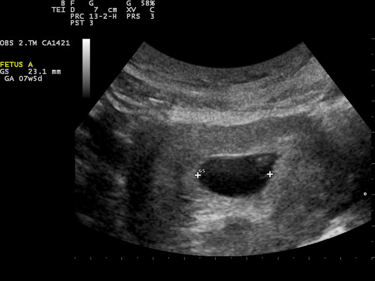
What if a patient could receive an ultrasound scan without a probe making contact with their skin? Engineers at MIT have developed a method that can accomplish this using laser. The new technique uses two lasers—that are safe on the skin and the eye—and can image the internals of a body remotely.
In a study, researchers from Massachusetts Institute of Technology(MIT) talk about the use of two lasers—one that when pointed at the body generates sound waves that bounce throughout the body, and the other that detects bounced waves—to create an ultrasound image similar to the ones produced using contact-based ones. These findings could greatly help in producing images of patients who cannot be probed through contact, such as burn victims, babies and people with sensitive skin.
Brian W Anthony, a senior author on the paper, said in a statement, "Imagine we get to a point where we can do everything ultrasound can do now, but at a distance. This gives you a whole new way of seeing organs inside the body and determining properties of deep tissue, without making contact with the patient."
Using lasers to generate and detect sound waves to form an image
The cutting-edge technology of photoacoustics employs the use of light aimed at the body at a particular wavelength to produce sound waves within it. The resulting vibrations due to light-induced sound waves are detected using transducers placed on the skin, which is converted into a photoacoustic image.

However, despite remotely using lasers to induce vibrations, transducers have to be in contact with the skin to detect them. Also, light cannot travel deep into the skin. Therefore, scientists have been able to image only blood vessels beneath the skin using photoacoustics, and not more.
The current study nevertheless manages to overcome the limitations of photoacoustic imaging. It addresses these limitations with the use of lasers to, both produce and detect sound waves. Lasers with a wavelength of 1,550-nanometers were chosen by the researchers. At this wavelength, light is easily absorbed by water—what the skin is composed of primarily—and by a large safety margin, is safe on the eyes and the skin.
How the new method works
A pulsed laser set at 1,550 nanometers is used to generate sound waves, while a continuous laser tuned at the same wavelength picks up these signals—basically acting as a motion detector. The frequency of the first laser changes as it is converted into sound waves, and is measurable. By mechanically scanning the lasers over the body, scientists can record data at different locations of the body and generate an image of the intended regions.
First, the method was tested by imaging metal objects enclosed within a gelatinous mould that mimics the composition of the skin. Next, they moved on to imaging animal skin, in this case, pigs. Finally, they moved on to imaging the forearms of healthy human beings using the technique.
They found that ultrasound image produced by laser was efficient in discerning finer features such as the borders between bones, muscles and fat. Another important discovery they made was that the laser-produced images were as accurate as conventional ultrasound imaging.
"It's like we're constantly yelling into the Grand Canyon while walking along the wall and listening at different locations," said Anthony. "That then gives you enough data to figure out the geometry of all the things inside that the waves bounced against—and the yelling is done with a flashlight," he added.
Possible commercial applications
The researchers intend to perfect the technique and better its performance by effecting improved discerning of tissue features. They also aim to improve the detection of the laser's capacity. However, what makes the new technique even more novel is the potential miniaturisation of the setup that can make ultrasound imaging portable and convenient.
Imagining the possibility of such a scenario, Anthony added, "When I get up in the morning, I can get an image of my thyroid or arteries, and can have in-home physiological imaging inside of my body."


