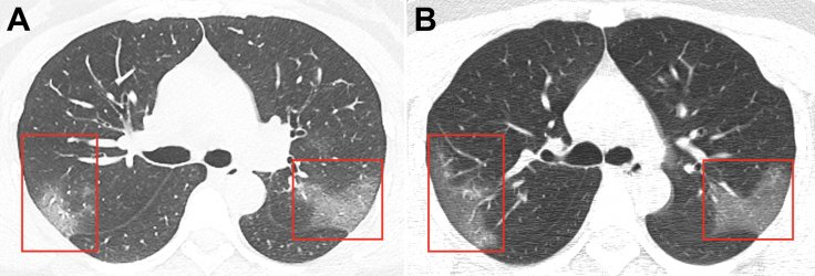
Chinese doctors have released images of the lungs of a 33-year-old woman who was diagnosed with the fast-spreading novel coronavirus or 2019-nCoV. The scans reveal the extent to which the coronavirus progressively affected her lungs.
The images that were released in a study, reveal the damaging speed at which the infection—that has afflicted nearly 15,000 and claimed 305 lives across 27 countries—advances.
Developed symptoms over five days
A resident of Wuhan, the woman sought medical assistance after a five-day ordeal of cough and fever brought on by an unknown cause. During admission, the woman revealed to the doctors that she had travelled to Lanzhou, China, six days prior to seeking treatment.
Her temperature was elevated—39C—when she reported to the hospital. Stethoscopic auscultation revealed that her breath was coarse in both the lungs and a series of laboratory tests indicated towards signs of an infection. After analysing her symptoms, laboratory results and CT scans, the doctors diagnosed her with 2019-nCoV.
CT scans reveal a stark picture
In the two comparative scan images released side-by-side, white patches can be seen in lower corners of both her lungs. In the report, the doctors describe these patches as "peripheral ground-glass opacities" as they seem to literally look like powdered glass. "What it represents is fluid in the lung spaces," said Paras Lakhani, a radiologist who examined the images but was not involved in the study.

The first picture titled 'A' shows a clear presence of white patches in both her lungs. However, in a juxtaposed picture titles 'B', growth in the size of the patches is clearly visible. According to the radiologist from Thomas Jefferson University, these white patches are characteristic of pneumonia triggered by different causes.
"You can see it with all types of infections — bacterial, viral, or sometimes even non-infectious causes," Lakhani told the Business Insider. "Even vaping could sometimes appear this way." However, white patches found in CT scans may not entirely aid in the diagnosis of the new coronavirus on its own as they can be misleading, said Lakhani.
More than what meets the eye
Generally, initial diagnosis based on the observation of white patches in CT scans may lead to doctors treating patients for pneumonia. "Typically, most hospitals will treat with antibiotics and patients will stabilize and then start to get better," added Lakhani.

However, this was not the case with the woman. After being treated for three days, and receiving inhalation of interferon-a protein used to treat certain infections—her condition continued to worsen. As the white patches grew progressively over three days, it helped doctors rule out the possibility of pneumonia as the condition is not known to progress so rapidly. "Pneumonia usually doesn't rapidly progress," said Lakhani.
Similar to other coronavirus infections
The researchers noted that the white patches extended up to the edges of the lungs, a feature similar to the ones found in the lung CTs of patients afflicted with other recent coronaviruses. "We saw that with Severe acute respiratory syndrome (SARS) and we saw that with the Middle East respiratory syndrome (MERS)," noted Lakhani.
He also noted that the scans of the lungs of the 33-year-old woman showed "a lot of similar features" as the other two coronaviruses. The SARS outbreak 0f 2002-2003 resulted in over 8,000 confirmed cases across 26 countries, while 2,494 confirmed cases of MERS have been reported across 27 countries as of November 2019.


